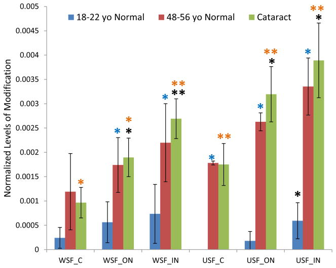
Figure 2.
GSH modification on Cys80 of βB1 crystallin in dissected lens regions separated into water soluble fraction (WSF) and urea soluble fraction (USF).* Indicates p< 0.05, ** indicates p< 0.005; Black asterisks show significant increasing modification compared with cortex in the same group of lenses. Blue asterisks show significant increasing of modification in middle-aged normal lenses compared with young lenses. Orange asterisks indicate significant increase in cataract lenses compared with young lenses. The difference of modification in cataract lenses compared with middle-aged normal lenses did not reach statistical significance.

