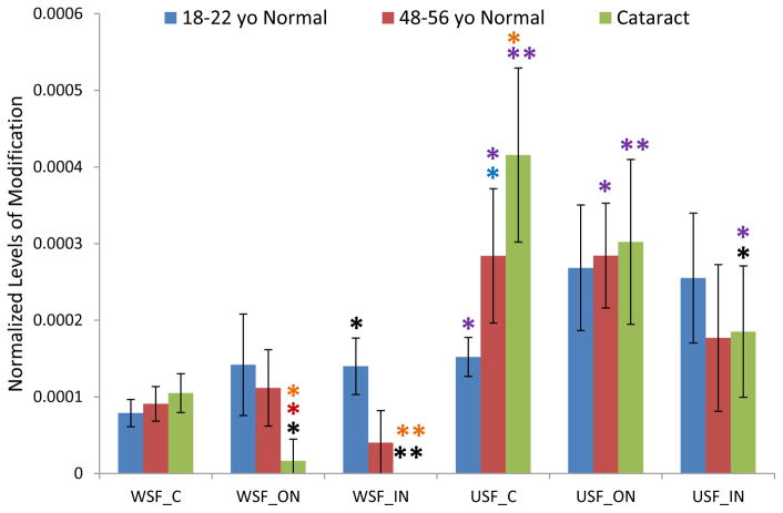
Figure 5.
GSH modification on Ser59 of αA crystallin in dissected lens regions separated into water soluble fraction (WSF) and urea soluble fraction (USF).* Indicates p< 0.05, ** indicates p< 0.005; Black asterisks show significant increasing or decreasing modification compared with cortex in the same group of lenses. Red asterisks show significant decreasing of GSH modification in cataract lenses compared with middle-aged normal lenses; Blue asterisks show significant increasing of modification in middle-aged normal lenses compared with young lenses. Purple asterisks indicate significant increasing in USF than in WSF.

