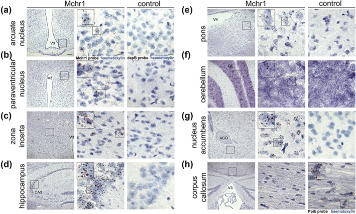
Figure 4.

In Situ Hybridization for Mchr1 in adult mouse brain. Representative images of brain sections from adult wild‐type mice following in situ hybridization using a Mchr1 specific probe (a‐h, left and middle columns; middle panel represents a magnified image of the annotated box, insets in the middle column are from individual cells in that panel). A dapB probe was used as a negative control (a‐g, right column) and a Ppib probe was used as a positive control probe (h, right). Neuroanatomical regions annotated include the third ventricle (V3), Cornu Ammonis 3 subfield of hippocampus (CA3), fourth ventricle (V4), granule layer (GL), anterior commissure (ACO), and lateral ventricle (LV). Probe staining in brown with haematoxylin counter stain. Scale bar in left column indicates 100 μm, scale bars in middle and right column indicate 10 μm. (n = 3)
