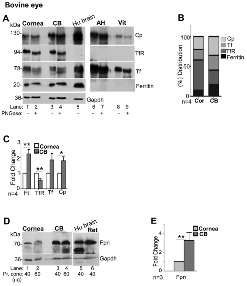
Fig. 3. Expression of iron modulating proteins in the cornea and ciliary body.

(A) Probing of lysates from bovine cornea (Cor) and CB for Cp, TfR, Tf, and ferritin shows the expression of all of these proteins in both samples (lanes 1–4). Deglycosylation results in faster migration of Cp, TfR, and Tf on SDS-PAGE, indicating the presence of glycans (lanes 2 & 4). AH and the vitreous show abundant presence of Cp and Tf, both of which migrate faster upon deglycosylation (lanes 7 & 9). No reactivity for TfR or ferritin is detected in these samples (lanes 6–9). Human brain lysate was processed in parallel as a positive control (lane 5). Gapdh served as a loading control. (Cor: cornea; Ft: ferritin). (B) Relative distribution of iron modulating proteins within each tissue shows higher expression of TfR relative to Cp and ferritin in the cornea, and higher levels of Cp relative to the TfR and ferritin in the CB. (C) Quantitative comparison of protein expression by densitometry shows significantly higher levels of ferritin and Cp, and lower levels of TfR in the CB relative to the cornea. All values were normalized to Gapdh that provided the loading control. Values represent fold change ± SEM of the indicated n. (D) Probing of Western blots of bovine cornea and CB for Fpn revealed increased expression of Fpn (3.2 fold) in CB relative to the cornea (lanes 1–4). Lysates from human brain and bovine retina were analyzed in parallel as controls (lanes 5 & 6). Gapdh served as a loading control. (E) Quantification by densitometry shows 3.2 fold higher levels of Fpn in the CB relative to the cornea. Values are mean + SEM of the indicated n. **p < 0.01. The full images of the cropped blots have been provided in the Supplementary Data.
