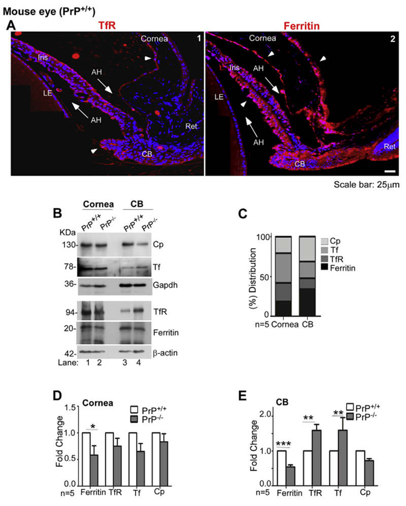
Fig. 7. PrPC modulates iron homeostasis in the anterior segment:

(A) Immunostaining of the anterior segment of PrP+/+ mouse eye for TfR shows a positive reaction in the CB, anterior surface of the iris, and corneal endothelium (Cor en) (panel 1). Reactivity for ferritin is more widespread, and is evident in the CB, iris, corneal endothelium, and corneal epithelium (Cor ep) (panel 2). Scale bar: 25 μm. (B) Probing of immunoblots from pooled lysates of cornea and CB of PrP+/+ and PrP−/− mice for Cp, Tf, TfR, and ferritin shows robust reaction for all proteins in both mouse lines (lanes 1–4). However, absence of PrP results in downregulation of ferritin in the cornea (lanes 1 & 2), and down-regulation of ferritin and Cp and upregulation of TfR in the CB (lanes 3 & 4). Gapdh and β-actin provide a loading control. The β-actin and Gapdh used in Fig. 7B is same as in Fig. 6B as the membranes were reprobed. (C) Percentage distribution of each protein in the cornea and CB shows relatively higher expression of Tf and TfR in the cornea, and Cp and ferritin in the CB. (D) & (E) Quantitation by densitometry following normalization with Gapdh and β-actin shows significant downregulation of ferritin in the cornea, and downregulation of ferrin and Cp and upregu; lation of TfR in the CB of PrP−/− mice relative to PrP+/+ controls. Values are mean ± SEM of the indicated n. *p < 0.05, **p < 0.01, ***p < 0.001. Full images of cropped blots are included in the Supplementary Data.
