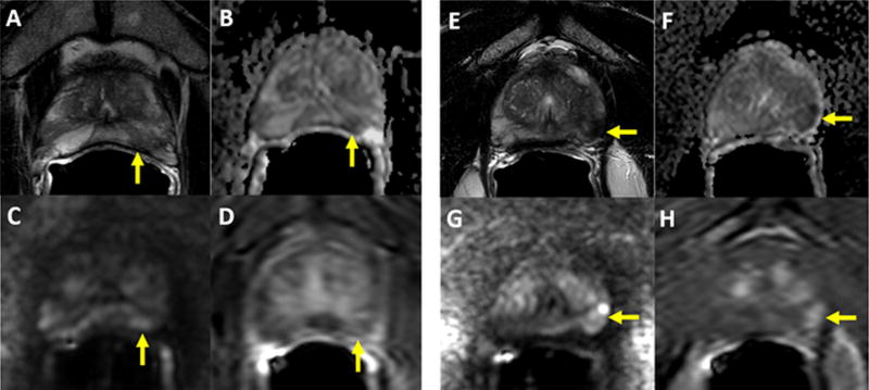
Figure 4.
Panels 4A–4D: 56-year old man with a serum PSA of 3.99ng/ml. Axial T2W MRI shows a heterogeneously hypointense lesion in the left apical PZ (arrow) (A), which shows diffusion restriction on ADC maps (B) and b2000 DW MRI (C) with relatively weak contrast enhancement on DCE MRI (D) (arrow). This lesion was scored as PIRADS 4/5 (T2=3, DWI=4, DCE=negative), whereas its in-house score was 2/5 since it is only positive on T2W and DW MRI. Targeted biopsy revealed benign prostate tissue within this lesion. Panels 4E–4H: 66-year old man with a serum PSA of 13.45ng/ml. Axial T2W MRI shows homogenously hypointense lesion in the left apical PZ with slight capsular bulge (arrow), which shows diffusion restriction on ADC maps (B) and b2000 DW MRI (C) with early contrast enhancement on DCE MRI (D) (arrow). This lesion was scored as PIRADS 4/5 (T2=3, DWI=4, DCE=negative), whereas its inhouse score was 4/5 since it is positive on T2W, DW MRI, DCE MRI with slight capsular bulge. Targeted biopsy revealed Gleason 3+4 prostate adenocarcinoma within this lesion.

