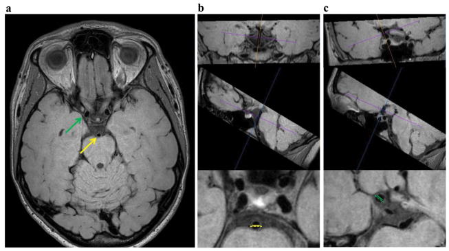
Figure 2.
Representative full FOV slice in the acquired plane (a) and example wall thickness measurements performed in a plane perpendicular to vessel course using a multi-planar reformatting tool (b,c). The basilar artery (yellow) wall thickness measured between the AICA and SCA (b) and supraclinoid ICA (green) wall thickness measurements (c) are demonstrated. The dashed lines represent the outer vessel wall diameter and the solid lines the luminal diameter.

