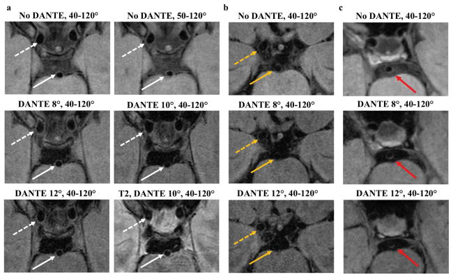
Figure 3.
Examples of the CSF suppression techniques in two subjects and one patient. Cropped views from a single slice in the acquired plane from the first subject (a) show improved CSF suppression in the prepontine cistern with DANTE. The walls of the basilar artery (white arrow) and supraclinoid internal carotid arteries (white dashed arrow) are well-depicted. A second subject (b) shows relatively good CSF suppression with the 3D TSE readout without DANTE (no DANTE, 40–120°) and therefore little further decrease in CSF signal with DANTE. In this subject, the DANTE 12° acquisition resulted in signal loss of the basilar artery vessel wall (dashed yellow arrow), making it hard to discern the wall throughout its circumference. The right ICA vessel wall (yellow arrow) remains well visualized with DANTE. Patient example (c) of 53-year-old female with multifocal intracranial atherosclerosis and prior left thalamic ischemic infarct. Hyperintense basilar artery wall lesion (red arrow) is more conspicuous with the addition of DANTE.

