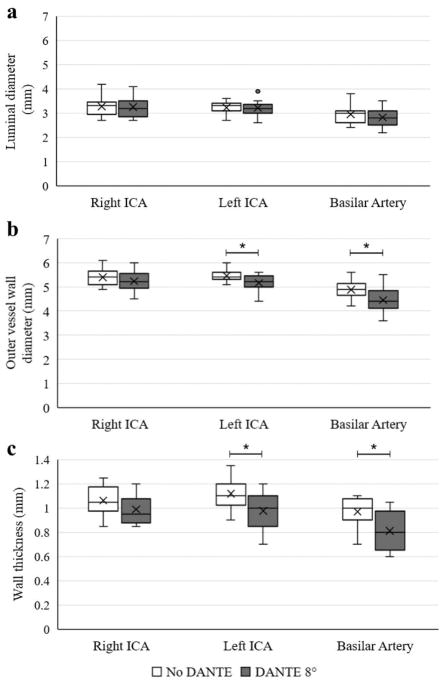
Figure 6.
Results of vessel wall measurements are shown by box plots of the luminal diameter (a), outer vessel wall diameter (b), and wall thickness (c) for the right ICA, left ICA, and basilar artery comparing the no DANTE, pulse sweep 40°–120° and the DANTE 8°, pulse sweep 40°–120° acquisitions. The central line represents the median, X the mean, the lower and upper margins of the box the 25–75th percentile, and the whiskers all data excluding outliers. *statistical significance with p<0.05.

