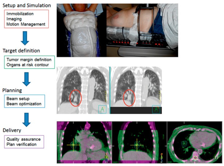
Figure 2. The Process of Stereotactic Body Radiation Therapy.
A) Two types of immobilization devices are shown. In the left panel, the patient is immobilized with BodyFIX. It utilizes vacuum technology to create a mold of the patient’s body contour. In the right panel, the patient is immobilized with a custom fitted foam frame with a paddle on the patient’s abdomen to minimize tumor motion with the respiratory cycle.
B) During simulation, a four-dimensional or respiratory CT scan is obtained. It allows for visualization of structures throughout the respiratory cycle. The scan on the left is during the exhale portion of respiration when the tumor motion is typically minimal. The right panel shows the maximum intensity projection (MIP) images. MIP is a surrogate for where the tumor is moving during respiration. The red oval encircles the tumor; the tumor appears fuller and larger on the MIP images.
C) Prior to delivering each fraction, a cone beam CT (CBCT) is performed on the treatment machine. The purple shade CT is the scan from the simulation. The green shade scan is from the CBCT prior to treatment. Depending on the location of the tumor, the CBCT can be matched to the simulation CT by bony landmarks or soft tissue to ensure the tumor is lining up properly prior to delivery of treatment.

