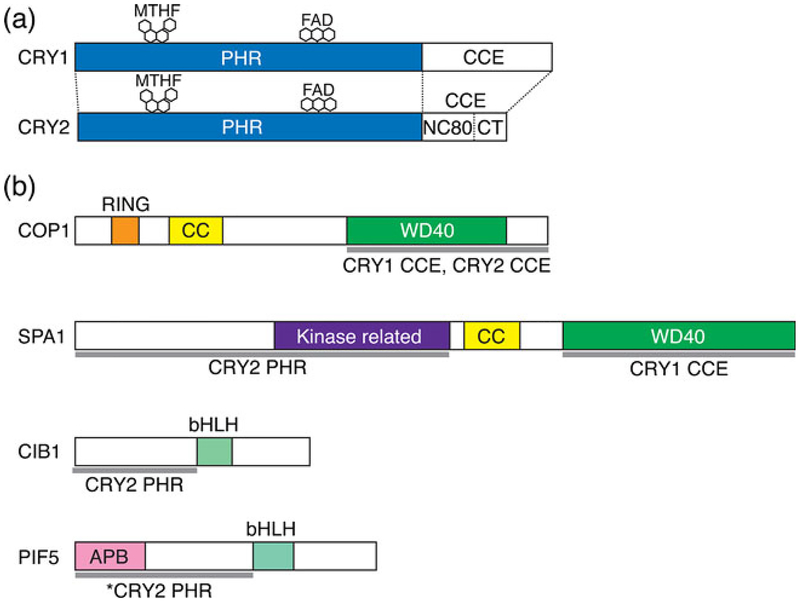
Figure 1.
The domain structure of CRYs and CRY-interacting proteins. (a) Schematic diagram showing domains in CRY1 and CRY2. MTHF, 5-methyltetrahydrofolate; FAD, flavin adenine dinucleotide; PHR, photolyase homology region domain; CCE, C-terminal extension domain; NC80, 80 residues without C-terminal tail in CCE domain; CT, C-terminal tail. (b) Schematic of CRY-interacting proteins showing the functional domains and CRY-binding sites. RING, RING finger domain; CC, coiled-coil domain; WD40, WD40 repeat; kinase-related, protein kinase-like domain; bHLH, basic helix–loop–helix domain, APB, active phytochrome B binding motif. The gray lines at the bottom of each diagram represent the CRY-binding regions. The binding domains of CRYs are also shown underneath each gray line. The asterisk indicates that CRY2 binds PIF5 with a mutation in the APB motif.

