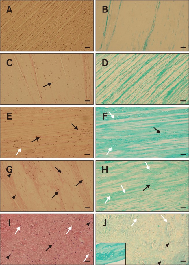
Fig. 1. Histological appearance of the cranial cruciate ligament with different structural changes. (A and B) Histological appearance of a normal cranial cruciate ligament showing typical fibroblast orientation with a spindle-shaped appearance, regular fascicular structure, and pale interfascicular areas. (C–F) Mild fibroblast transformation of the nuclei into an ovoid to round shape, some columnar chondroid transformation (black arrows) and formation of perinuclear halo (white arrows), loss of even distribution of the fibroblasts within the ligament, regular fascicular structure, and increased mucopolysaccharide content (alcian blue/periodic acid-Schiff [AC-PAS] stain). (G–J) Significant chondroid metaplasia with the formation of columns (black arrows) and clones of transformed fibroblasts (black arrowheads and inset in panel J), irregular fascicular network, and a significant increase in mucopolysaccharides (AC-PAS). White arrows, perinuclear halo. H&E stain (A, C, E, G, and I). AC-PAS stain (B, D, F, H, and J). Scale bars = 100 µm (A–J). 400× (inset in panel J).