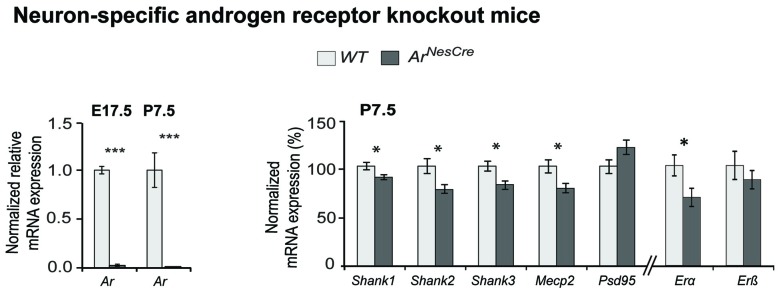
Figure 3.
Comparative Shank gene expression analysis by nCounter in the cortex of wild-type (WT) and ArNesCre mice. (Left) Loss of androgen receptor (Ar) expression in cortical neurons was confirmed by qPCR. The deletion of Ar-exon2 was shown on mRNA level in cortical tissue. (Right) Gene expression analysis in cortex of WT and ArNesCre mice at P7.5. The loss of Ar resulted in a decreased expression of the Shank and Mecp2 genes (n = 7 WT and seven KO mice, two male and five female animals in each group). The analysis could not be stratified by sex due to low numbers. Gene expression was normalized against four reference genes (Gapdh, Hprt1, Hspd1 and Sdha). Error bars indicate SEM (unpaired two-tailed Student’s t-tests, *P ≤ 0.05; ***P ≤ 0.001).

