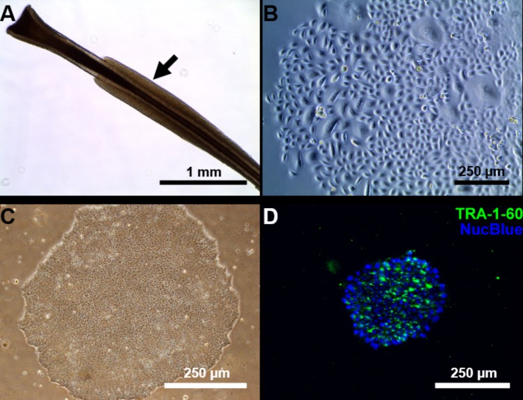
FIGURE 1.
Reprogramming of follicular keratinocytes. (A) Plucked human hair follicle from the scalp region. The hair follicle is in the anagen phase. A visible outer root sheath (black arrow) is highly visible and intact, thus providing a higher number of keratinocytes. (B) Confluent keratinocyte culture isolated from hair follicles, at 12 days of culture. The cells display typical keratinocyte morphology (strongly adherent cells with cobblestone appearance). (C) Representative Sendai viral generated iPSC colonies. Passage 1 iPSC culture manually picked from emerging colony in Cytotune 2.0 transfected keratinocytes. The well-defined borders of the colony are, characteristic of iPSC colonies. (D) The generated iPSC culture stains positively for TRA-1-60 (green), a known pluripotency marker. Nucleus is stained with NucBlue (blue).

