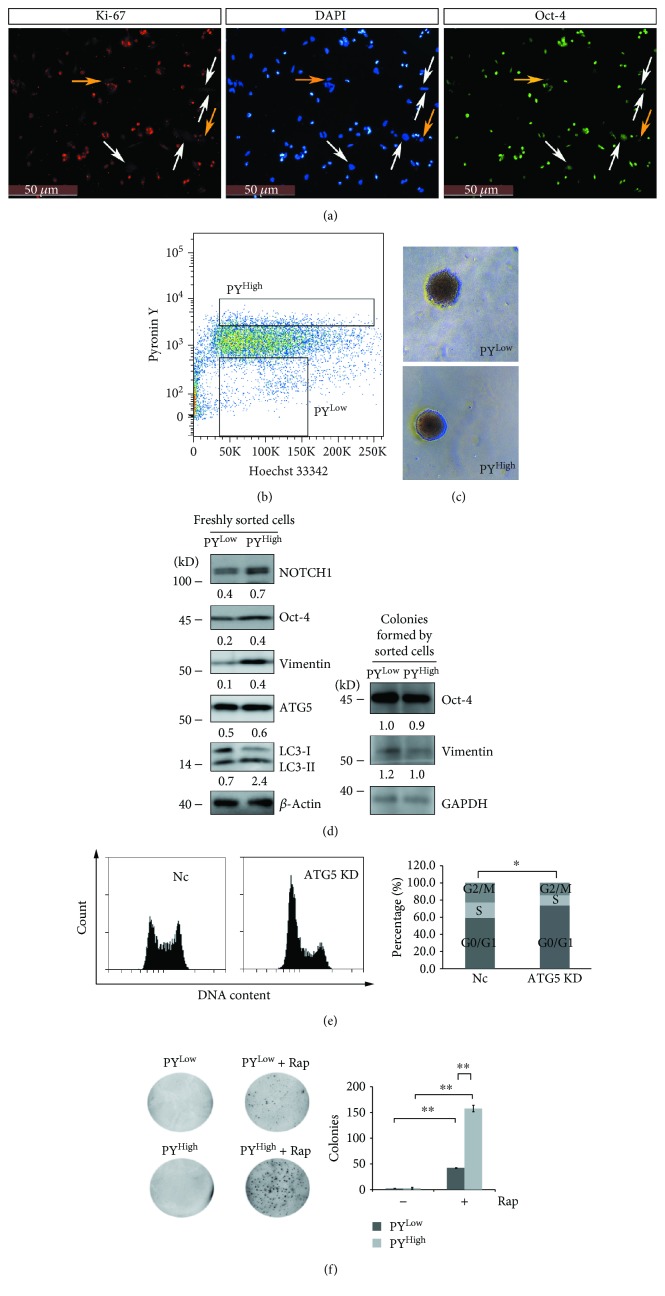
Figure 4.
Autophagy promotes quiescent ovarian cancer spheroid cells to reenter the cell cycle. (a) Immunostaining of Oct-4 and Ki-67 in A2780 spheroid cells. Cells were cultured under spheroid culture condition for 48 h, trypsinized, and seeded back to attach to the plate for 4 h, fixed, stained with anti-Oct-4 antibody (green) and anti-Ki-67 antibody (red), counterstained with DAPI (blue), and observed with a fluorescence microscope. (b) Cell sorting of PYLow and PYHigh cells. A2780 spheroid cells were trypsinized, stained with Hoechst 33342 and pyronin Y, and sorted by flow cytometry. (c) Colony formation of sorted PYLow and PYHigh cells on soft agar. Sorted PYLow and PYHigh cells described in (b) were grown on soft agar for 28 d to form colonies. (d) Western blot analysis of the protein levels of Oct-4, NOTCH1, vimentin, ATG5, and LC3-I/II from extracts of freshly sorted PYLow and PYHigh cells or colonies formed by sorted PYLow and PYHigh cells. The colonies described in (c) were collected, resolved in SDS loading buffer, and subjected to Western blot analysis. The relative intensity of indicated proteins normalized to housekeeping protein was shown. LC3-II/LC3-I ratios were calculated. (e) Cell cycle analysis of Nc and ATG5 shRNA A2780 cell strains. The cells were trypsinized, fixed, digested with RNase A, stained with propidium iodide, and subjected to cell cytometry analysis (mean ± SEM, n = 3). (f) Colony formation of PYLow and PYHigh cells with or without rapamycin (Rap, 1 μM) on soft agar. 1.5 × 10⁴ of PYLow and PYHigh cells were cultured on soft agar with or without Rap for 14 d. The images were taken with a gel imaging system, and the colonies were counted with ImageJ software (mean ± SEM, n = 2).

