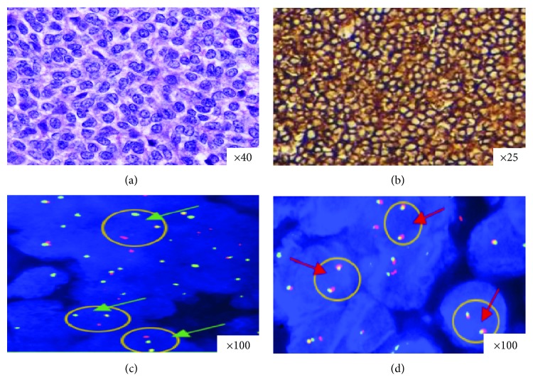
Figure 1.
H&E staining, CD99 immunostaining, and dual-color FISH for the EWSR1 gene in selected EPS cases. (a) H&E stained section showing diffuse proliferation of round blue cells. (b) Diffuse, intense, and membrane expression of CD99 antibody. (c) Dual-color FISH performed with break-apart EWSR1 probes reveals nuclei in which one pair of the probe signals is split apart due to a rearrangement in the EWSR1 gene (green arrows). Red arrows indicate a two-signal pattern in normal cells (d).

