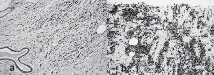
Fig. 3.
Histopathological photomicrographs. a Photomicrography of the mass revealing small cells with little cytoplasm. Alveolar-type spaces are present containing desquamated small, round, and poorly differentiated skeletal muscle cells (hematoxylin and eosin, ×40). b Photomicrograph of immunohistochemical evaluation of the primary mass showing diffusely positive staining for myogenin (×20).

