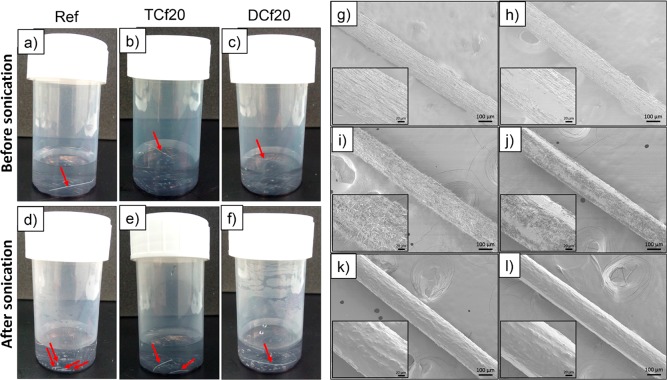
Figure 9.
Photographs of unmodified and organosilane-modified CNF filaments in deionized water (a–c) before and (d–e) after sonication. SEM micrographs at 100× magnification of (g and h) ref, (i and j) TCf20, and (k and l) DCf20 (g, i, and k) before and (h, j, and l) after rubbing (see Table 1 for nomenclature). The insets are micrographs at 500× magnification. The DC-modified filament remained intact after sonication and sunk to the bottom of the container (noted with the arrow added in part f). In contrast, the unmodified filament was fragmented upon sonication (noted by the arrows added in part d).

