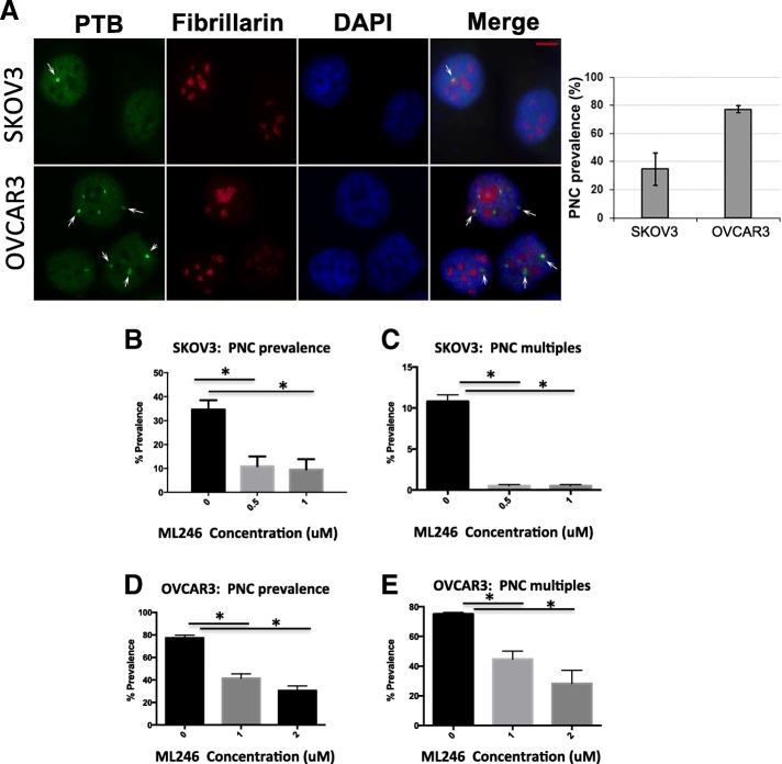
Fig. 1.
PNC prevalence in ovarian cancer cell lines. a Immunofluorescent staining was done in SKOV3 and OVCAR3 for PTB, a component of PNC (green, marked by arrows) that are located adjacent to nucleoli (fibrillarin staining in red) within the nucleus (DAPI staining in blue). Bar = 5 μm. b and c SKOV3 and d and e OVCAR3 cells were treated with ML246 at 0.05uM, 1uM or 2 uM or DMSO for 24 h and % PNC prevalence as well as cells carrying multiple PNCs was calculated. * p < 0.05

