
Fig. 3.
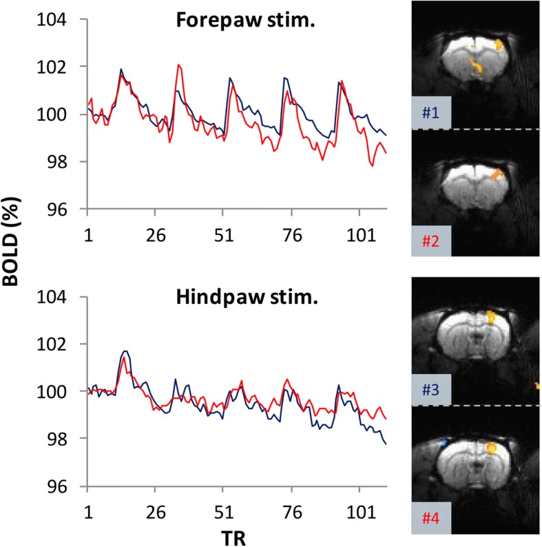
Represented results of fMRI checkpoints before the formal study. Before the fMRI investigation of the brain responses to SNI nerve transection, we used the S1 BOLD response to forepaw stimulation to confirm whether the experimental conditions were suitable for fMRI scanning (upper panel). Subsequently, we used the S1 BOLD response to hind paw stimulation to verify whether the sciatic nerve was intact after implantation of the nerve transection device (lower panel). In the represented data, the S1 forepaw and hind paw areas exhibited consistent spatial and temporal responses to forepaw and hind paw stimulation, respectively, among the successive scans. #1–4 indicate the scan order. TR stands for the repetition time for each acquisition, which was 2 s in the current study