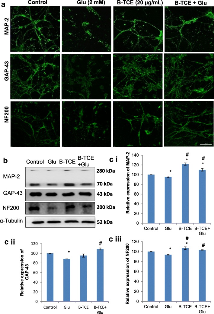
Fig. 2.
B-TCE pretreatment suppressed the neuronal degeneration induced by glutamate by modulating the expression of neuronal structural markers. a) Confocal micrographs of Control, Glutamate (2 mM), B-TCE (20 μg/mL) and B-TCE + Glu treated primary cerebellar neurons immunostained for MAP-2, GAP-43 and NF200. b) Representative immunoblots for MAP-2, GAP-43 and NF200 where α-Tubulin was used as internal control. c) Histogram representing relative expression of MAP-2 (i), GAP-43 (ii) and NF200 (iii) obtained from normalized relative optical densities of bands. Data was compared between Control and other groups such as Glutamate, B-TCE and B-TCE + Glu (*p ≤ 0.01) as well as Glutamate alone with B-TCE and B-TCE + Glu groups (#p ≤ 0.01). Confocal Images were captured at 60X objective (Scale bar: 50 μm)

