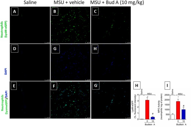
FIGURE 4.

Fifteen hours after the injection of MSU crystals, knee joint wash of LysM-eGFP mice was collected for quantification of fluorescence intensity [Original magnification 20×, panels (A-G)] using a confocal microscope. Percentage of fluorescence is represented in panel (H). Knee joint of Swiss mice was dissected for determination of neutrophil recruitment assessed by determination of MPO activity (I) by a colorimetric method. ∗p < 0.05 vs. saline group; #p < 0.05 vs. 0 mg/kg group, one-way ANOVA followed by Tukey’s post hoc.
