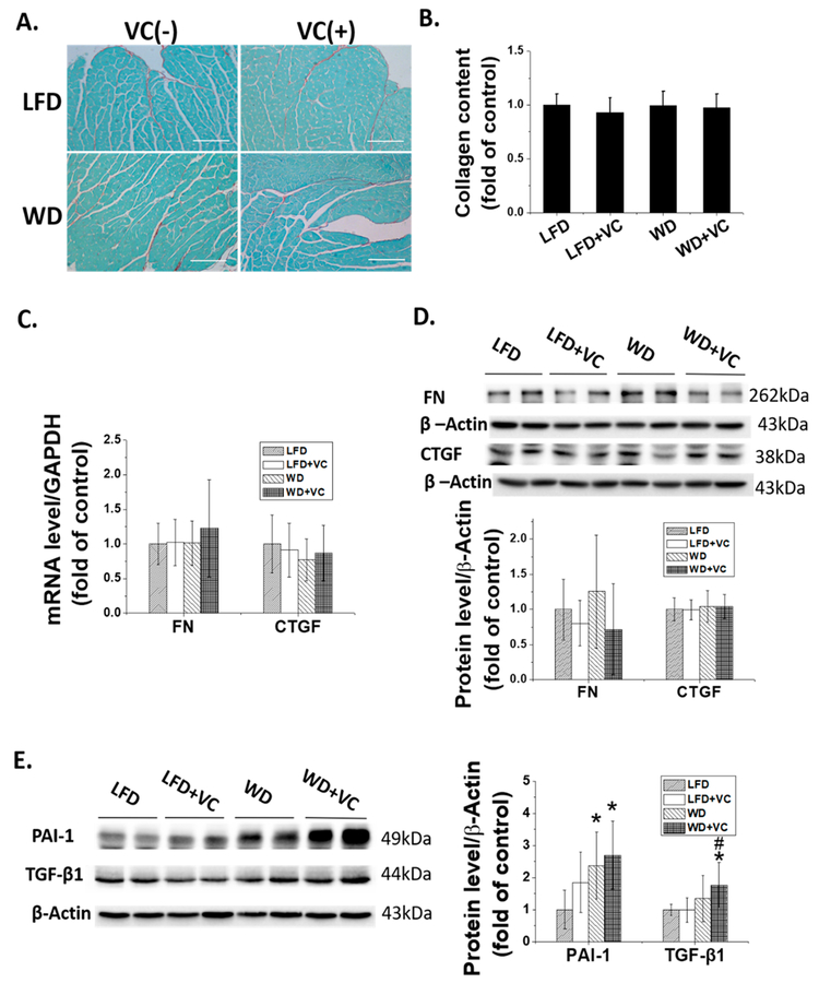
Figure 3.
WD and VC did not cause apparent cardiac fibrosis but increase the expression of some profibrotic cytokines. (A) Cardiac fibrosis, determined by Sirius Red staining of collagen accumulation (collagen is red; 200×, scale bar = 100 μm), and (B) quantitative analysis of Sirius Red staining for collagen accumulation. (C) mRNA expression of CTGF and FN was determined by RT-qPCR. Protein expression of (D) CTGF and Fibronectin, (E) PAI-1 and TGF-β1 were determined by Western blot. ∗, versus LFD group, P < 0.05; $, versus LFD+VC group, P < 0.05.

