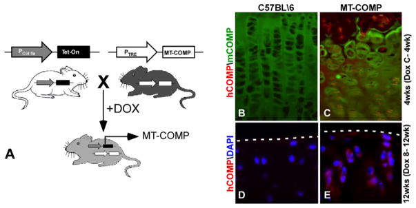
Fig. 1.
MT-COMP transgenic mouse. (A) MT-COMP bigenic mouse line was generated using the Tet-On system with the tetracycline responsive element (TRE) driving human D469del-MT-COMP expression. The type II collagen promoter (Col IIa) drives rtTA (Tet-On) expression in chondrocytes. (B) In the presence of doxycycline (DOX), the bigenic MT-COMP mouse expresses mutant human D469del-COMP in growth plate and articular chondrocytes as visualized using human-specific COMP antibody. MT-COMP (hCOMP in red) expression is detected in the murine growth plate chondrocytes at 4 weeks with administration of DOX from conception to 4 weeks (C-4wk) (C), whereas mouse COMP (mCOMP in green) is primarily extracellular (B and C). Similarly, MT-COMP is found in articular cartilage chondrocytes of 12 week mice administered DOX from 8 to 12 weeks (E) but not in controls (D). The articular cartilage border is marked by a dashed line. DAPI staining of nuclei are shown in blue (D and E).

