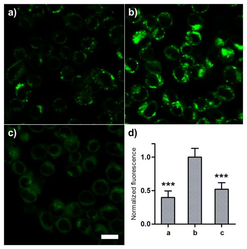
Figure 2. Compound 7 stains macrophages undergoing phagosomal acidification.
RAW264.7 cells (preincubated or not with zymosan) were treated with 7 (100 nM) for 15 min and imaged by confocal microscopy. Fluorescence staining of 7 in (a) nonactivated macrophages, (b) zymosan-activated macrophages, and (c) zymosan-activated macrophages treated with bafilomycin A (100 nM); (d) quantification of fluorescence emission represented as means (n = 4) and error bars as SD, *** for p < 0.005 compared to b. Scale bar: 20 μm.

