Figure 2.
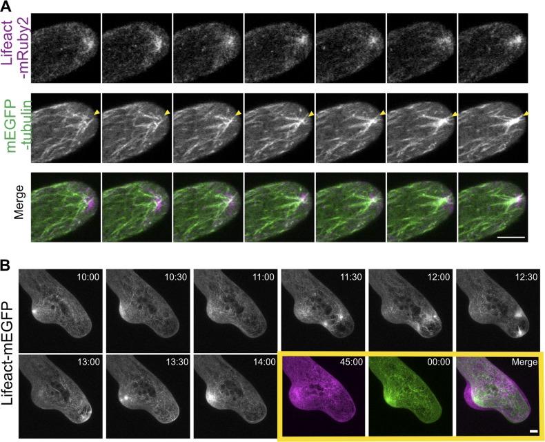
Microtubules are required for maintaining directional growth and the formation of the actin cluster near the cell apex. (A) A WT cell stably expressing mEGFP-tubulin (green) and Lifeact-mRuby2 (magenta). Microtubules coalesce within the actin cluster (yellow arrowheads). Images are single focal planes acquired every 2 s. (B) Actin cytoskeleton was perturbed when microtubules were disrupted. A WT cell expressing Lifeact-mEGFP growing in the presence of 10 µM oryzalin. In the yellow box, the first and the last image from the time lapse movie were merged to show cell expansion. Time stamps represent min:s. Images are maximum projections of z-stacks acquired every 30 s. Bars, 5 µm. See also Videos 3 and 4.

