Figure 6.
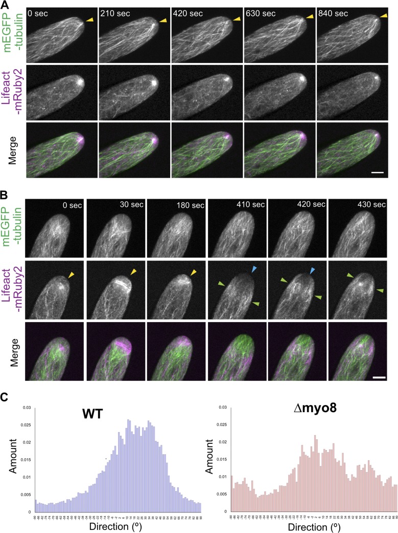
Myosin VIII is required for maintaining both apical actin and microtubule structures. (A) A WT cell stably expressing mEGFP-tubulin (green) and Lifeact-mRuby2 (magenta). Images are maximum projections of z-stacks acquired every 15 s. (B) A Δmyo8 cell expressing mEGFP-tubulin (green) and Lifeact-mRuby2 (magenta). Yellow arrowheads, abnormal apical actin accumulation. Blue arrowheads, very low actin abundance near the cell apex. Green arrowheads, waves of actin bundles migrating throughout the cell body. Images are maximum projections of z-stacks acquired every 10 s. Bars, 5 µm. See also Video 8. (C) Microtubule orientations are quantified from selected frames in A and B using the ImageJ plugin Directionality. The histograms represent the number of structures within an image in a given orientation.

