. 2018 Oct 2;7:e38856. doi: 10.7554/eLife.38856

Figure 1. Overexpression of S.aureus gpsB inhibits cell division in B. subtilis.

(A) Luria-Bertani agar plates streaked with wild type B. subtilis (WT, strain PY79), or otherwise wild type B. subtilis harboring an inducible copy of gpsBBs (GG18), gpsBBs-gfp (GG19), gpsBSa (GG7), or gpsBSa-gfp (GG8) integrated into the chromosome, in the absence (left) or presence (right) of inducer. (B–M) Morphology of cells of different deletion mutants of B. subtilisezrA, GG35; ΔponA, CS26; ΔprkC, CS24; ΔgpsB, CS40; ΔdivIVA, CS94) harboring an inducible copy of gpsBSa grown in the absence (B, D, F; H, J, L) or presence (C, E, G, I, K, M) of inducer. (N–O) Localization of FtsZ-GFP in a strain (GG9) harboring an inducible copy of gpsBSa grown in the absence (N) or presence (O) of inducer. Membranes (red; B–O) visualized using the fluorescent dye FM4-64; chromosomes (blue; B–M) visualized using DAPI; FtsZ-GFP localization (green; N–O). Scale bar: 1 μm. Genotypes are listed in Key Resources Table.

Figure 1.

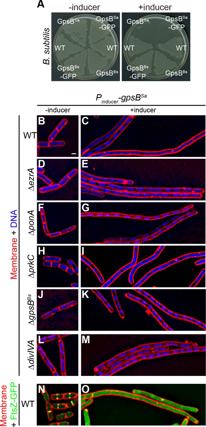
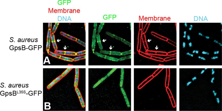
Figure 1—figure supplement 1. GpsB sequence and subcellular distribution in S.aureus.

Figure 1—figure supplement 1.

(A) Alignment of amino acid sequences of GpsB from various strains. Identical residues are highlighted in gray; Leu35, (site of loss-of-function suppressor mutation in gpsBSa) is boxed; Ala23 (which corresponds to Leu24 in the L. monocytogenes ortholog and mediates membrane association) is denoted with an asterisk. (B) Immunoblot of cell extracts from B. subtilis cells (WT; PY79) or cells harboring an inducible copy of gpsBSa (GG7) in the absence (-) or presence (+) of inducer using rabbit antisera raised against purified GpsBSa or SigA (loading control). Note: anti-GpsBSa antiserum does not detect B. subtilis GpsB. (C) Immunoblot of cell extracts from S. aureus cells (WT; SH1000) or cells harboring a plasmid-encoded inducible copy of gpsBSa (SH1000 pPE45) in the absence (-) or presence (+) of inducer using rabbit antisera raised against purified GpsBSa or B. subtilis SigA (which also recognizes S. aureus SigA). (D) Circular dichroism spectra of purified GpsBSa (black) or GpsBSa-L35S (red). (E) Total extracts (T) of S. aureus cells producing GpsB-GFP or GpsBL35S-GFP were prepared and fractionated into soluble supernatant (S) and insoluble pellet (P) fractions by ultracentrifugation. GpsB-GFP was detected using rabbit anti-GpsB antiserum raised against purified GFP mut2. (F) Immunoblot of cell extracts from S. aureus cells harboring an inducible copy of gpsB antisense RNA (left) or empty vector (right) grown in the presence of inducer using antisera recognizing GpsB or SigA.
Figure 1—figure supplement 2. GpsBSa-GFP localizes to division septa in B.subtilis.

Figure 1—figure supplement 2.

Localization of (A) GpsBSa-GFP or (B) GpsBSa-L35S-GFP in B. subtilis. First panel: overlay of GFP, membrane visualized using FM4-64, and DNA visualized using DAPI; second panel: GFP fluorescence; third panel: membrane; fourth panel: DNA. Arrows indicate sites of cell division.