. 2018 Oct 2;7:e39213. doi: 10.7554/eLife.39213

Figure 8. Accessory subunits of complex I.

Central subunits (see Figure 4) are shown in grey, accessory subunits are labelled and coloured. (A) Side views, (B) view from the matrix (left) and from the intermembrane space (right) with peripheral arm subunits removed for clarity.

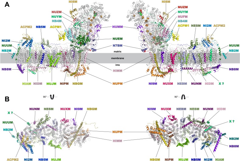
Figure 8.

Figure 8—figure supplement 1. Assignment of accessory subunits.

Figure 8—figure supplement 1.

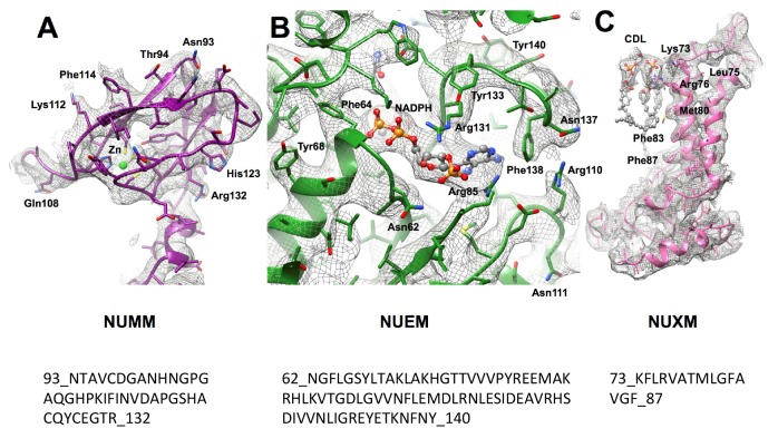
Example densities with corresponding sequence stretches are shown for the Zn binding domain of NUMM and for the NADPH binding domain of NUEM. In agreement with secondary structure predictions subunit NUXM has four αhelices, two of which are transmembrane. Side chains were modelled for short sequence stretch in this subunit.
Figure 8—figure supplement 2. Assignment of accessory subunits.

Figure 8—figure supplement 2.

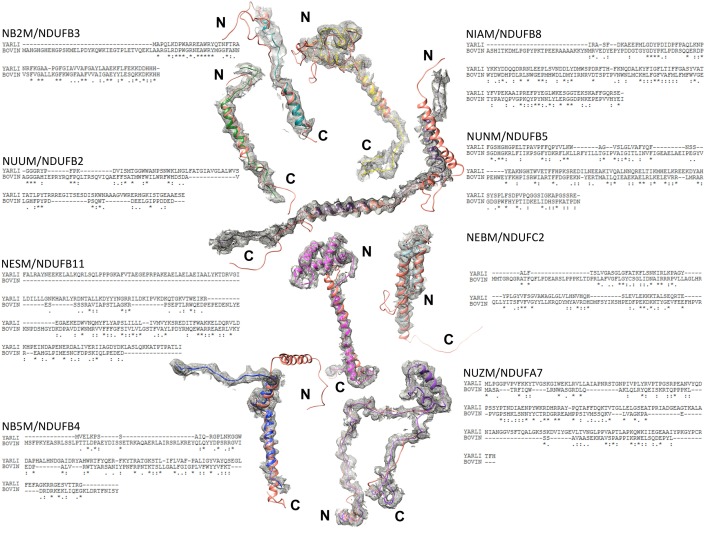
Sequence and structural alignments with bovine complex I (PDB ID 5O31 (Blaza et al., 2018), bovine subunits, orange) are shown for eight subunits that were modelled largely or entirely as poly-alanine.