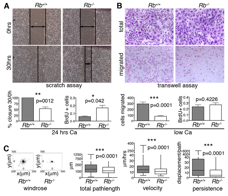
Figure 2. Rb null keratinocytes have motility defects.
(A) Epithelial cell layers were generated by culturing wildtype and Rb-/- keratinocytes in high calcium for 24 hours. The Rb-/- keratinocytes had an impaired ability to repair scratches, as indicated by representative images and quantification (left graph; n=6 lines), and displayed ectopic proliferation, as assessed by BrdU incorporation (right graph; 3 lines/genotype, ~2000 cells. (B) Single Rb-/- keratinocytes (cultured in low calcium) showed impaired migration 12 hrs post-plating in Boyden chambers relative to wildtype controls, as judged by crystal violet staining of cells with representative images above and quantification in left graph (n=6 lines/genotype, plated in triplicate), without proliferation changes, which were assessed by BrdU incorporation (right graph; n=3 lines/genotype, ~500 cells). (C) Time-lapse microscopy was conducted for 15 hrs to track the path of individual wildtype and Rb-/- keratinocytes (n=3 lines and >350 cells per genotype). From left to right: windrose plots and graphs show that Rb-/- cells had significant reduced total pathlength, velocity, and persistence compared to their wildtype counterparts. Statistical significance was determined by Student’s t-test.

