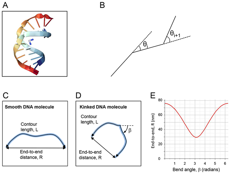
Figure 1.

(A) Crystal structure of duplex DNA (rainbow color) containing a cisplatin 1,2-d(GpG) intrastrand cross-link (blue & white molecule), inducing a bend angle of 35° to 40° and local unwinding of the double helix by 25° 37. (B) Schematic depiction of the DNA simulations. (C, D) Schematic depiction of an unbent DNA fragment (C) and a DNA fragment with a bend, β (D). (E) Plot of equation (2); end-to-end distance, R, as a function of the bend angle, β, for a 300 bp DNA fragment.
