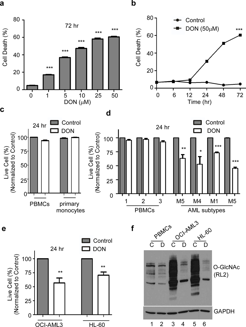
Figure 2. Blocking protein O-GlcNAcylation kills AML cells.

DON treatment blocks O-GlcNAcylation and subsequent cell death in OCI-AML3 cells was monitored (a) in a dose-dependent manner after 72 hr treatment and (b) in a time-dependent manner with DON (50 μM) treatment. (c) Cell viability of normal PBMCs and primary monocytes 24 hr after DON (50 μM) treatment compared to the untreated control. (d) Cell viability of PBMCs and AML patient blast samples treated with DON or untreated control after 24 hr. (e) Effect of DON (50 μM) on the cell viability of OCI-AML3 and HL-60 cells after 24 hr treatment. (f) Western blot showing O-GlcNAc profile of PBMCs, OCI-AML3 and HL-60 using O-GlcNAc (RL2) antibody. Cells were incubated (16 hr) as indicated. C-untreated control or D-DON (50μM). Actin was used as an endogenous loading control. Statistical significance was calculated using unpaired Student’s t-test. N=3; *p<0.05, **p<0.01, ***p<0.001.
