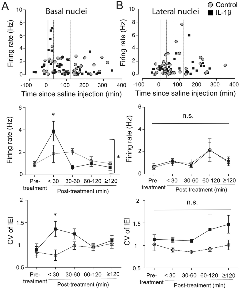
Figure 5: IL-1β preferentially impacted firing of neurons in the basal nucleus.
(A) Basal nucleus (BA) of the BLA complex. Firing rate of individual neurons from the BA is plotted against time (upper). The firing rate between the two groups was compared among five different time windows (middle; pre-treatment (baseline), <30 min, 30–60 min, 60–120 min, and ≥120 min post-treatment). There was a significant interaction between treatment and time (middle; p=0.014, two-way ANOVA). CV of IEI was significantly increased at <30 min window in IL-1β group (lower). *p<0.05 by Holm-Sidak’s post hoc test after significance in two-way ANOVA. (B) Lateral nucleus (LAT) of the BLA complex. Firing rate of individual neurons from the lateral nucleus is plotted against time (upper). The firing rate between the two groups was compared among the same five different time windows. There was no significant interaction (p=0.992, twoway ANOVA) and no significant main effects. There was a main effect of treatment on CV of IEI (p=0.003, two-way ANOVA), though no single IL-1β treatment time point emerged as significant compared to control in post hoc tests (p>0.05, Holm-Sidak’s post hoc test). The line through 0 time-point represents time of treatment. Dotted lines represent 30 min, 60 min and 120 min post-treatment. Plots show mean ± S.E.M.

