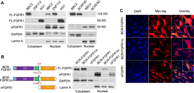
Figure 4. Cleavage and subcellular localization of the chimeric FGFR1 kinases.

Cell fractionation studies using SCLL cell lines (A) show the relative locations of the various chimeric kinases between the cytoplasm and nucleus. The relatively pure isolation of both cell compartments is shown by the presence of cytoplasmic (GAPDH) and nuclear (LaminA) markers. In the three examples, the 55 kD truncated derivative (nFGFR1) can be seen exclusively in the nucleus. Similarly, the presence of the nuclear truncated form is seen in the AML-derived BCRF8C cell line compared with murine BBC2, both expressing BCR-FGFR1. Schematic representation of BCR-FGFR1 showing the coiled coil domain (CC), the serine/threonine kinase domain (STK), the Dbl homology domain (DH) and the FGFR1 tyrosine kinase domain (TK). The arrow indicates granzyme B cleavage site (GrB) at Asp432 of FGFR1 (B, left). The absence of the truncated nuclear variant in transfected 3T3 cells following mutation of the GrB cleavage site (BCR-FGFR1m) is seen compared with the parent chimeric kinase (B, right). The presence of the exogenous truncated form of FGFR1 is shown exclusively in the nucleus. Immunofluorescence analysis of the three variant kinases further demonstrated the cytoplasmic location of the full length BCR-FGFR1 chimeric kinases as well as exclusive nuclear presence of the truncated form in the NIH 3T3 cells (C). Insets represent the cell indicated by the arrow at higher magnification.
