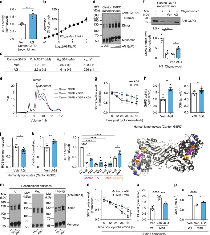
Fig. 3.
AG1 (activator of G6PD) induces biochemical changes in the Canton variant. a Increased activity of Canton G6PD enzyme by AG1 (n = 5, ***p = 0.0002, two-tailed unpaired Student’s t-test) and b a dose response curve of AG1. c AG1 changed kinetic parameters of Canton G6PD. d AG1 promoted dimerization of Canton G6PD (n = 3). e Size-exclusion FPLC (calibrated Superdex 75 10/300 GL column) profile of purified Canton G6PD in the presence of G6P or AG1. f AG1 reduced proteolytic susceptibility of Canton G6PD (n = 3, ***p < 0.001, *p < 0.05, one-way ANOVA). The protein levels were normalized to the non-treated (NT) enzyme level. g Cycloheximide-chase assay using lymphocytes carrying the Canton variant (n = 3). Protein levels were normalized to the level of each enzyme at time 0 h. h, i, j AG1 increased G6PD activity in cell lysates with the Canton variant (n = 4, **p = 0.0032, two-tailed unpaired Student’s t-test), mildly enhanced a GSH level (n = 7, *p = 0.0282, two-tailed unpaired Student’s t-test) and reduced a ROS level in culture (n = 6, *p = 0.0452, two-tailed unpaired Student’s t-test). k AG1 increased viability of lymphocytes carrying the Canton variant (n = 6, **p = 0.003, two-tailed unpaired Student’s t-test). l, m AG1 activated other major G6PD variants, including A− (V68M, N126D; blue spheres), Mediterranean (S188F, orange spheres), and Kaiping (R463H, yellow spheres) variants, respectively (n = 4, ****p < 0.0001, **p < 0.01, *p = 0.011, one-way ANOVA), and promoted their dimerization (n = 3). Purple spheres in the structure represent the side chain of R459. n Cycloheximide-chase assay using fibroblasts carrying the Mediterranean variant (n = 4, *p = 0.0437, two-tailed unpaired Student’s t-test). o, p AG1 significantly decreased a ROS level (n = 6, ***p = 0.0001, ****p < 0.0001, one-way ANOVA) and increased a GSH level in those cultures (n = 3, *p = 0.0214, ****p < 0.0001, one-way ANOVA). 100 μM and 1 μM of AG1 were used for in vitro assays and cell-based assays, respectively. 5% DMSO (stock) was used as vehicle. For FPLC assay, 500 μM AG1, 200 μg of Canton G6PD recombinant enzyme and 10 mM G6P were used. Cells were subjected to serum starvation for 48 h. Error bars represent mean ± SEM. MW: molecular weight, FPLC: fast protein liquid chromatography, NT: no treatment, Veh: vehicle, WT: wild-type, Med: Mediterranean fibroblast

