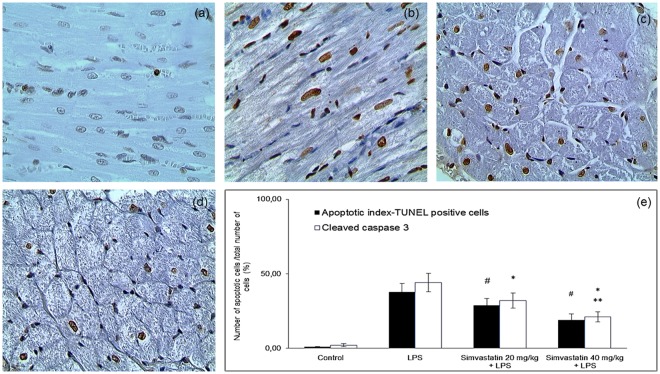
Figure 3.
Simvastatin inhibited apoptosis in rat myocardial tissue in LPS induced inflammation detected by TUNEL staining, magnification 400x. Brown stained nuclei indicate TUNEL-positive cardiomyocytes. The apoptosis increased significantly in the LPS (b) and simvastatin group (c) compared with the control group (a). Note that induction of sepsis by LPS resulted in a marked appearance of TUNEL-positive cardiomyocytes (arrow) quantified and shown as AIs (black columns) (e), which was significantly reduced by simvastatin 20 (c) and simvastatin 40 (D). (e) Quantitative analysis of apoptotic cells counted in immunohistochemically stained myocardial sections for cleaved caspase 3 and corresponding frequencies of TUNEL positive cardiomyocytes are shown, *p < 0.01 in comparation with LPS group, **p < 0.05 in comparation with simvastatin 20, #p < 0.05 in comparation with LPS group.

