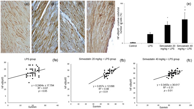
Figure 6.
Simvastatin increased NF-κB expression in rat myocardial tissue in LPS induced inflammation. Representative images with semi-quantitative analysis of survivin positive cells in myocardium in groups that were challenged with LPS or either pretreated with simvastatin 20 or simvastatin 40 before LPS. Immunohistological staining of myocardial tissues was performed using a p65-specific antibody to evaluate NF-κB p65 expression in cardiomyocytes, magnification 200x and 400x. (a) Control group. Note subsets of cardiomyocytes positive for NF-κB/p65 in the cell cytoplasm and/or nucleus in the LPS group (b), and intensive nuclear immunostaining in the simvastatin 20 (c) and simvastatin 40 (d) groups. (e) Semiquantitative analysis of NF-κB/p65 expression. *p < 0.01 in comparation with LPS group, #p < 0.05 in comparation with simvastatin 20 group. (f) The correlations of survivin expression and NF-κB/p65 positive cardiomyocytes within the experimental groups (fa, fb, fc).

