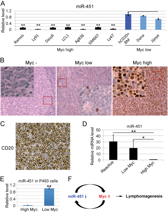
Fig. 7.
miR-451 expression is disrupted in human B lymphoma cells with hyper Myc activity. a qRT-PCR analyzing the level of miR-451 in Myc highly-expressed and Myc-low-expressed human lymphoma cell lines with human CD20 positive bone marrow lymphocytes as control. The Y-axis shows the relative amount of miR-451. Representative study from three different experiments is shown. b Immunohistochemical staining of representative specimens from human diffuse large B cell lymphomas (DLBCL) patients showing the high Myc and low Myc protein levels. Inflammatory reactive lymph nodes with undetectable level of Myc as negative control (Myc-). Insets (400×) represent tissues in red squares. c Immunohistochemical staining of CD20 showing the identity of B cell lineage. d q-RT-PCR analyzing miR-451 levels in human DLBCL samples with high and low Myc protein levels. RNA from reactive lymph nodes with undetectable Myc used as control. The Y-axis shows the relative amount of miR-451. q-RT-PCR was repeated twice. e q-RT-PCR analyzing miR-451 levels in subcutaneously transplanted human lymphoma P493 cells with a Myc-inducible system. The Y-axis shows the relative levels of miR-451. f Model for the interaction of miR-451-Myc: decrease of miR-451 expression derepresses Myc in B-lymphocytes and high Myc level inversely inhibits miR-451 transcription. This positive feedback loop sustains the Myc level above the threshold that contributes to B-lymphomagenesis. (*) p < 0.05 (t-test), (**) p < 0.01 (t-test)

