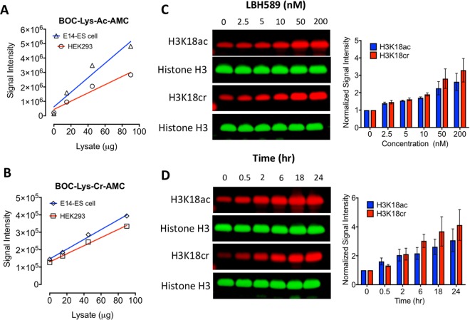
Figure 2.
HDAC inhibition increases both H3K18ac and H3K18cr in a dose-dependent manner. (A) Deacetylase and (B) decrotonylase activities were measured using increasing concentrations of whole-cell extracts from mouse ES and HEK-293T cells using Boc-Lys(Ac)-AMC and BOC-Lys(Cr)-AMC substrates. Average plots of n = 3 technical replicates. ES cells were treated with either, increasing concentrations of LBH589 for 24 hrs (C), or treated with 50 nM of LBH589 for indicated time (D), before histones were extracted and subjected to quantitative western blotting using an Odyssey scanner. Levels of H3K18ac and H3K18cr were normalized to the level of histone H3 and graphs show the average normalized signal intensity (mean ± SEM; n ≥ 3). Uncropped scans of western blot gels are in Supplementary Fig. 2A,B.

