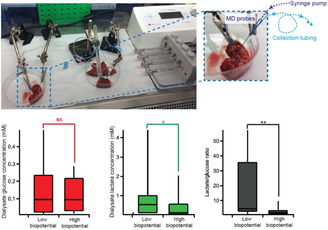
Figure 5.
Laboratory setup for dialysate collection. (a) Two probes are inserted into non-cancerous tissue (left) and cancerous tissue (right) and perfused simultaneously using a syringe pump. The dialysate is collected in 0.75 m lengths of collection tubing. (b) Box and whisker diagrams comparing dialysate glucose levels (red), lactate levels (green) and lactate/glucose ratio (black) for paired omentum tissue with low and high biopotential (n = 12). Statistics tested using two-tail Wilcoxon sign rank test.

