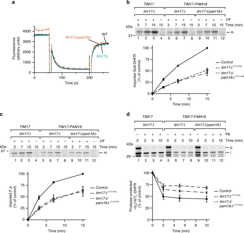
Fig. 2.
Mitochondria containing a Tim17–Pam18 fusion display mild-matrix import defects. a Membrane potential (Δψ) of mitochondria was assessed using the Δψ-sensitive dye DiSC3(5). Fluorescence quenching by mitochondria was recorded before and after addition of valinomycin. b, c [35S]-labeled precursor proteins were imported into the mitochondria and import stopped after indicated time points using antimycin A, valinomycin, and oligomycin (AVO). Samples were Proteinase K (PK)-treated and analyzed by SDS-PAGE and digital autoradiography. Results are presented as mean ± SEM, n = 3. The longest import time of the WT sample was set to 100%. m, mature protein; tim17∆+T17-P18 tim17∆ + TIM17–PAM18. d The inward-driving force of the import motor was assessed using [35S]-labeled b2(167)∆-DHFR in the presence of methotrexate (MTX). After 15 min import, Δψ was dissipated using valinomycin. In a second step, the precursor was chased for indicated time before PK addition. The amount of processed intermediate was quantified (100%: amount of processed intermediate without protease treatment). Results are shown as mean ± SEM, n = 4. p, precursor; i, intermediate

