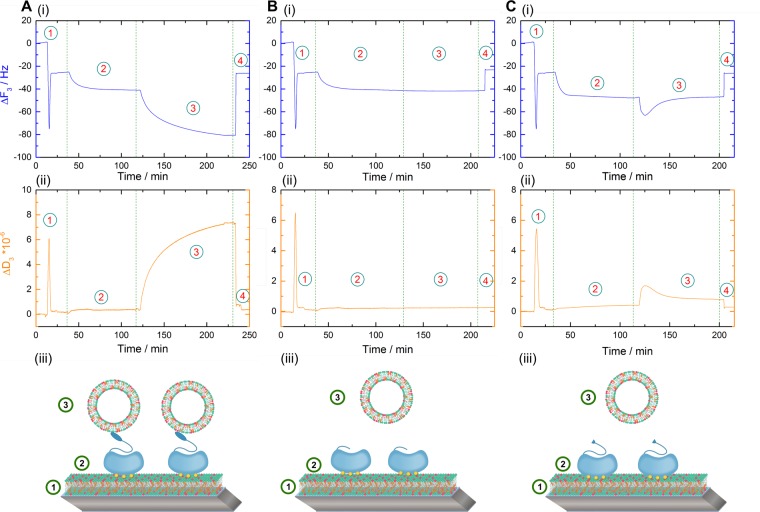
Figure 3.
Time resolved frequency (i) and dissipation (ii) monitoring of the Ca2+ dependent and reversible lipid binding and membrane bridging/linking by WT AnxA2 (A) and the N-terminal deletion mutants AnxA2 Δ 32 (B) and AnxA2 Δ14 (C) in the QCM-D setup. Numbers illustrate the sequential addition of lipids and proteins: Step 1, SLB formation by SUV adsorption and rupture; step 2, adsorption of the respective AnxA2 derivative; step 3, secondary vesicle application in the form of LUVs; step 4, release of all Ca2+-dependently bound material by chelation with EGTA. Models representing the respective experimental setup are shown below the QCM-D recordings (iii). Shown are representative examples of typical experiments carried out n = 12 independent times with at least three different protein batches and vesicle preparations.

