Figure 5.
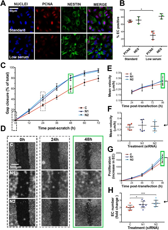
Nestin siRNA knockdown increases EC proliferation in vitro. (A) Immunofluorescence staining of nestin and PCNA in HUVEC cultured in standard or low serum medium (B) Quantification of nestin or PCNA positive cells (n = 3, 34–73 cells analysed/experiment) Paired t-test *p-value < 0.05. (C) HUVEC were treated with control (‘C’) or one of 2 anti-nestin (‘N1’ and ‘N2’) siRNAs and cultured to confluence for 48 h, before a ‘wound’ was generated and gap closure was measured at 12 h intervals (means ± SEM, n = 13). (D) Representative phase contrast images were captured at 0 h, 24 h and 48 h post wound creation (48 h, 72 h and 96 h post-transfection, respectively). HUVEC were seeded to 50% confluence and transfected with C, N1 or N2 siRNA, subsequently (E) migration velocity (n = 6), and (G) cell proliferation (n = 8) were measured every 24 h for 96 h. Individual data points from 96 h post-transfection are shown for velocity and migration (F and H, respectively). Green boxes indicate measurements taken at the same time point post-transfection (96 h). (All graphs: mean ± SD, significance calculated by one-way ANOVA).

