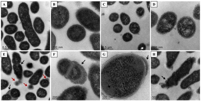
Figure 6.
Cellular ultrastructure observation. (A,B) Phenotypes of wild-type strain Yb2. (A) Integral and regularly shaped cells, a few cells were in division; (B) regularly shaped cells at higher magnification. (C,D) Phenotypes of complemented strain cYb2ΔbioF. (C) Integral and regularly shaped cells; (D) regularly shaped cells at higher magnification. (E–H) Phenotypes of mutant strain Yb2ΔbioF. (E) Dropped vegetations (red arrows) and most cells were in division (black arrows); (F) relatively thickened cell walls (black arrow); (G) partially thickened cell walls (black arrow); (H) irregular shapes (black arrow).

