Figure 1.
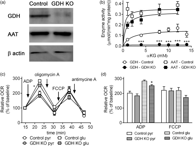
Expression and enzyme activity of GDH and AAT and respiration of isolated brain mitochondria. The expression of GDH and AAT determined by Western blot (loading control: β-actin) (a) and enzyme activities of GDH and AAT (b) in mitochondria from control (white) and GDH KO mice (black). The respiration of mitochondria from control (white) and GDH KO mice (gray) was measured in the presence of pyruvate + malate or glutamate + malate as substrates (c, d). The relative change of OCR was calculated by normalization of the 3rd basal measurements to 100% (control: 72.3 ± 17.4 pmol/min; GDH KO: 61.9 ± 10.6 pmol/min with pyruvate + malate; control: 57.6 ± 12.8 pmol/min, GDH KO: 45.8 ± 8.3 pmol/min with glutamate + malate) (c, d). The data shown are mean ± SEM of values obtained on 3 independent mitochondria preparations (each preparation and condition: 13–15 wells/experiment).

