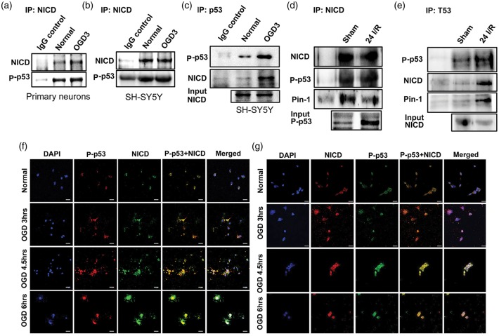
Figure 4.
NICD associates with p53. (a–e), Representative immunoblots of NICD and p53 co-immunoprecipitate in primary cortical neurons (a) and a neuroblastoma SH-SY5Y cell line (b and c) following 3 h of OGD and in ischemic brain 24 h following ischemia and reperfusion (I/R) or sham surgery (d and e). Confocal immunofluorescence images of primary cortical neurons (f) and neuroblastoma SH-SY5Y cells (g), show staining of NICD (f, green; g, Red), P-p53 (f, red; g, green), and nuclear marker 4′6-diamidino-2-phenylindol (DAPI) (blue). Both NICD and P-p53 staining are increased and co-localized at the indicated OGD time-points. Scale bar: 20 µm.

