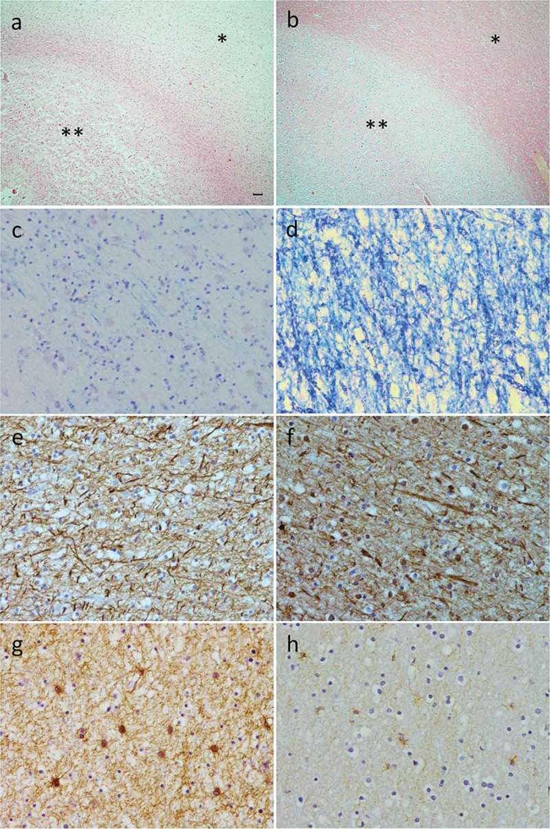
Figure 5.

Histological changes in a brain region with severe WMH (right frontal pole; a, c, e, and g) and in a region with no WMH (right occipital lobe; b, d, f and h). Hematoxylin and eosin staining demonstrates severe grey matter spongiosis (**) and pallor of the white matter (*) in the frontal pole (a). In the occipital lobe (b) there is intact cytoarchitecture of the grey matter (**) and white matter (*). Luxol fast blue staining demonstrates extensive myelin pallor in the white matter of the frontal pole (c) but intact myelin in the occipital lobe (d). SMI31 immunohistochemistry demonstrates relative preservation of axons in the white matter of both frontal pole (e) and occipital lobe (f). Severe gliosis was present in frontal pole white matter (g) but only mild in occipital lobe white matter (h). Scale bar in (a) represents 100µm for (a) and (b), and 10µm for (c) to (h).
