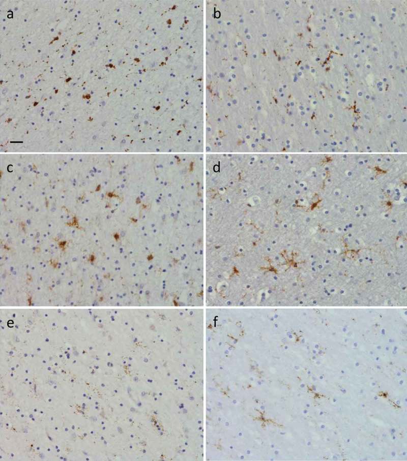
Figure 6.

Presence of microglia in right frontal pole white matter (a), (c) and (e), where there were severe WMH, and right occipital white matter (b), (d) and (f), where WMH were absent. Images show results of immunohistochemistry for CD68 (a) and (b), CR3/43 (c) and (d) and Iba1 (e) and (f) positive microglia. There were more amoeboid microglia in the frontal versus occipital white matter detected using CD68 (a) versus (b), and CR3/43 (c) versus (d). Iba1 positive microglia were ramified in occipital white matter (f) but few in number and severely dystrophic in frontal white matter (e). Scale bar in (a) represents 20µm in all panels.
