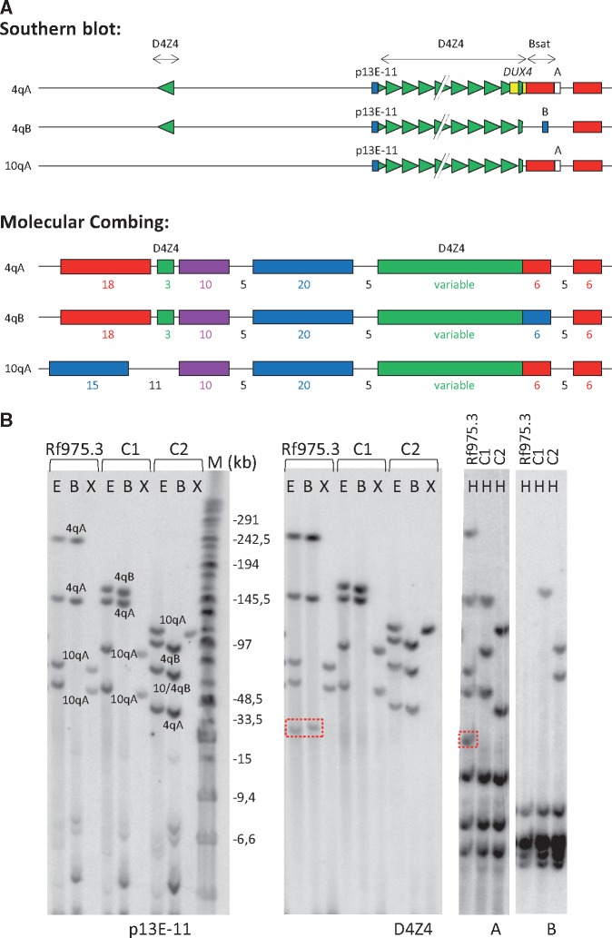
Figure 2.
(A) Schematic representation of D4Z4 locus on chromosomes 4qA, 4qB and 10qA for both Southern blot (top) and molecular combing (bottom) analysis. For both methods, D4Z4 units and the highly homologous inverted D4Z4 repeat unit on chromosomes 4qA and 4qB are indicated in green. For Southern blot analysis, the position of probes p13E-11 (blue), D4Z4 (green), A (white) and B (blue) are indicated, as well as position of the DUX4 gene (yellow). For molecular combing, the position and colour of the fluorescence probe which generate the unique genetic Morse code for each chromosome are indicated with sizes in kb. (B) Representative images of the Southern blot analysis in FSHD2 patient Rf975.3 and two unrelated controls (C1 and C2). Rf975.3 carries a D4Z4 duplication fragment, not visible by probe p13E-11, resistant to BlnI and hybridizing to the D4Z4 and A probes (hence 4qA-derived) (boxed repeat fragment). For the left two blots, DNA was digested by EcoRI and HindIII (E); EcoRI and BlnI (B) and XapI (X) and hybridized with probes p13E-11 and D4Z4, respectively. On the right two blots, DNA was digested with HindIII (H) and hybridized with probes A and B, respectively. Marker lane (M) in kb is indicated.

