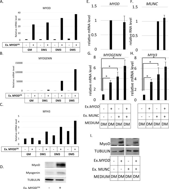
FIG 6.
Expression of MYOD partially rescued the MYOD−/− cell phenotype but did not significantly stimulate the induction of MYOGENIN or MYH3 by MUNC. (A to C) qRT-PCR analysis of MYOD−/− cells with or without doxycycline-mediated overexpression of exogenous MYOD in proliferating (GM) and differentiating (DM1, DM3, and DM5) cells as indicated on the x axes. Levels of expression were measured for MYOD (A), MYOGENIN (B), and MYH3 (C) mRNAs and normalized to GAPDH and are shown relative to MYOD−/− cells without overexpressed MYOD in GM. (D) Western blot showing exogenous MyoD and myogenin proteins induced in MYOD−/− cells when exogenous MyoD protein is induced by doxycycline. Tubulin was used as a loading control. (E to H) qRT-PCR analysis of MYOD−/− cells stably overexpressing MUNC, transiently overexpressing exogenous MYOD, and differentiated for 2 days. Levels of expression were measured for MYOD (E), MUNC (F), MYOGENIN (G), and MYH3 (H) mRNAs. The data were normalized to the GAPDH expression level and are shown relative to control cells. The values represent three biological replicates and are presented as means and SEM. Statistical significance was calculated using a Wilcoxon-Mann-Whitney test. *, P < 0.05. (I) Western blot analysis showing induction of exogenous MyoD protein in MYOD−/− cells when transiently transfected with MYOD. Tubulin was used as a loading control.

